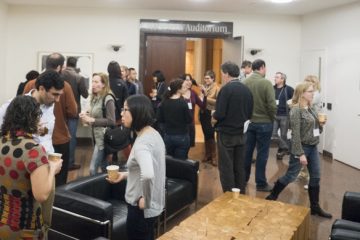
The 31st Annual Spring Workshops were held on May 1st, 2014 at the Marine Biological Laboratory in Woods Hole, MA. Three workshops ran concurrently throughout the afternoon, and covered topics ranging from computational image analysis with ImageJ to STM/AFM microscopy techniques and cathodoluminescence on geological samples.
[divider scroll_text=”Return to Top”]
[space height=”4″]
Introduction to Scanning Probe Microscopy Workshop
In this workshop, the attendees were introduced to the basic principles and applications of Scanning Probe Microscopy (SPM). In the first half, Brent LaPointe from Nanosurf Inc. gave a presentation on how this group of imaging techniques work and how they are employed in various scientific and technical fields. After the coffee break, the attendees had a chance to concentrate on two of the most popular SPM techniques, namely the Atomic Force Microscopy (AFM) and Scanning Tunneling Microscopy (STM). Fettah Kosar from Harvard gave a short demonstration on the Nanosurf NaioSTM small desktop system by imaging the surface of a HOPG (Highly Oriented Pyrolytic Graphite) at atomic resolution. Then Brent LaPointe demonstrated the capabilities of the Nanosurf FlexAFM system by imaging a sample provided by Stephen Senft from MBL, which contained sections of the chromatophores (color-changing organelles) from a squid. The level of interest and engagement by all the four attendees were indicated by their questions and comments throughout the session.
Fettah Kosar
Immediate Past President
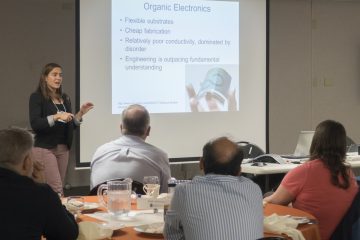
Digital Image Analysis with ImageJ
The “Digital Image Analysis with ImageJ” workshop was taught by Dr. Lai Ding and Daniel Tom of the Enhanced Neuroimaging Core at Harvard University. ImageJ and its more actively developed derivative,Fiji, are open source image processing packages that have grown a vast user base in the biological and physical sciences. Dr. Ding, a veteran user of ImageJ, presented a neatly organized and engaging overview of ImageJ’s most powerful functions. Appealing to both beginner and intermediate users, Dr. Ding’s workshop covered everything from image compression and file types to image segmentation. Most of the workshop was performed as hands-on demonstrations and allowed the workshop participants a chance to test the functions for themselves. The overview of ImageJ culminated with a peak into the versatile ImageJ macro language. Workshop participants left with the ability to perform batch operations to their images and make thousands of measurements all with the click of the “Run” button. If you missed Dr. Ding’s NESM workshop, consider attending one of his multi-day workshops at Harvard University. For more information, visit: http://www.neurodiscovery.
Blair Rossetti
President
[divider scroll_text=”Return to Top”]
[space height=”4″]
Cathodoluminescence Imaging – Maximizing Compositional Analysis in Mineralogy: A practical guide to commercial and exploration mineralogy techniques, and how they can be supported with selected CL imaging.
The cathodoluminescence workshop went off quite well, with a lively talk about the use of CL imaging, which uses the emission of visible wavelength light under electron bombardment in many crystalline materials, to materials investigations in many fields, but with an emphasis on mineralogical applications. The attendees were able to learn about the fundamentals of the dynamics of electron induced luminescence, as well as the ways in which this illumination allows for rapid material identification based on color, as well as quantitative analysis by interpretation of the emission with a monochrometer.In addition to examining a wide variety of polished mineral samples from Alaska, Brazil, California, and East Africa, we were able to look at granite samples from nearby Carlisle, MA. Applications of this technique to industrial materials science applications were also overviewed, using synthetic crystals manufactured locally. Comparison of the CL effects on samples was also made using optical petrography and SEM-EDS analysis thanks to the use of the Wood’s Hole MBL central microscopy facility laboratory. All in all, the workshop covered a lot of ground, allowed for some dynamic question and answer time, and introduced an underused technique to the suite of tools presented at the Spring Symposium.
Jared Kelly
Clerk
0 Comments