This year’s spring symposium was held on May 2, 2014 at the Marine Biological Laboratory in Woods Hole, MA. The symposium was outstanding in its variety of speakers and the depth of
their presentations.
Zvonimir Dogic, Physics Department at Brandeis University: Dr. Dogic showed us the beauty of self-assembling “model membrane” systems made from phage viruses. The phage viruses are used as rod-like colloidal particles that bind to one another and form phase-separated regions of virus, which separate from the aqueous buffer background. The effect is similar to oil drops in water – except the “oil drops” are made from viruses. Using DIC and Phase-contrast microscopy, these solid phases were clearly distinguishable from the buffer background. Using fluorescence and polarization microscopy, the tilt, orientation, and angle of the viruses within the phase-separated region could be directly visualized. All imaging was performed with various forms of light microscopy, showcasing the broad applicability of light microscopy to tackle this problem at the interface of biology and materials science.
Wei Guo, Photonics Laboratory at UMass Lowell: Dr. Guo uses electron and light microscopy to study novel light-emitting materials that could be the next generation of LED lighting systems. Our insatiable craving for power is dominated by our need to light up the night and 22% of all energy use is due to light bulbs of some kind. Thus, making more energy efficient lights could have significant energy-saving consequences. Growing and characterizing gallium-nitride and indium-nitride heterogeneous structures can control the wavelengths to give brighter, whiter light. Dr. Guo fabricates bamboo-like nanowires of repeating intervals of GaN and InN, and characterizes their abilities using SEM and fluorescence imaging. Guo uses microscopic characterizations of these nanostructures to help solve the energy crisis – one nanowire at a time.
Ilke Arslan, Pacific Northwest National Lab: Dr. Arslan is working on cutting-edge methods that use electron microscopy to visualize three dimensional materials systems and their dynamics over time. By tilting the sample in a scanning transmission electron microscope, Arslan is able to make 3D reconstructions of materials over time. By shaping the material to better fit into the TEM holder, even greater angles can be achieved, which results in more detailed 3D information. In addition to 3D tomography, Arslan is performing chemical reactions and observing the effects on the material in the microscope in real-time. By pushing the boundaries of how we can see, Arslan is learning new information about the microstructure of materials in true four-dimensions – space and time.
Jennifer Morgan, Marine Biology Laboratory: Dr. Morgan is using electron microscopy to study the molecular basis of Parkinson’s Disease. Interestingly, Morgan is using an unexpected model system – the lamprey – to study this disease. Lamprey is a good model system because, although more simple than human anatomy, they have a true brain and spinal cord which allows direct imaging the synapses of the motor neurons. The synapse is where the chemical communication between neurons and between neurons and muscles, through small neurotransmitter-containing organelles called synaptic vesicles. The lab studies the synuclein proteins that are associated with Parksinson’s Disease and which might play a role in synaptic vesicle trafficking. Morgan is able examine the effects of missing or mutations of this protein on the synapse structure and number of vesicles at the synapse for signaling using transmission electron microscopy of thin sections of fixed and embedded tissues. Morgan has found that when there is an overabundance of the synuclein protein (as there is in some cases of genetic Parkinson’s Disease) , there are fewer vesicles and more plasma membrane at the synapse, which suggests that synaptic vesicle endocytosis is somehow being impaired. Through direct microscopy imaging and the use of a unique and useful model system, Morgan is learning about the molecular mechanisms behind this debilitating neurological disease.
Amy Gladfelter, Biology Department at Dartmouth College: Dr. Gladfelter is using numerous microscopic techniques in to discover the molecular and cellular mechanisms of the little-studied septin proteins. Septins are a cytoskeletal filament that interact with the cell membrane to help organize cellular compartments and communicate with the rest of the cell. Teaming up with groups at the Marine Biological Laboratory at Woods Hole, Dr. Gladfelter has employed Fluorescence Correlation Spectroscopy (FCS), Total Internal Reflection Fluorescence (TIRF) Microscopy, polarized fluorescence, polarized TIRF, and multifocus polarization techniques to study the problems using both cellular and in vitro reconstitution experiments. She has discovered that septin filaments are apolar and often elongate through the annealing of shorter filaments. The orientation of the membrane-bound filaments is controlled in cells and serves to connect with other cytoskeletal elements. Through her multi-method, multi-scale approach that utilizes multiple microscopy methods, Gladfelter is learning how septins function in the cell.
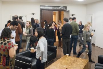
In addition to an excellent assortment of talks, the Symposium featured its annual exhibiting vendor session. Representatives of fifteen of NESM’s wonderful corporate sponsors manned tables, gave demonstrations and spoke with meeting attendees about all the latest in microscopy-related technology, equipment, and innovations. It was a treat to get a chance to engage with the vendors and discover firsthand all of the great developments in our field.
Jennifer Ross & Rylie Walsh
President-Elect & Corresponding Secretary

0 Comments